
Japanese scientists have found a way to observe coagulation activity in the blood as happened, without the need for invasive procedures.
Using a new type of microscope and artificial intelligence, scientists have shown how plattttiness “agglomeration” can be followed in patients with coronary disease, opening the door to safer and more personalized treatment.
Plates are tiny blood cells that rush to connect the cut skin when we suffer from a cut.
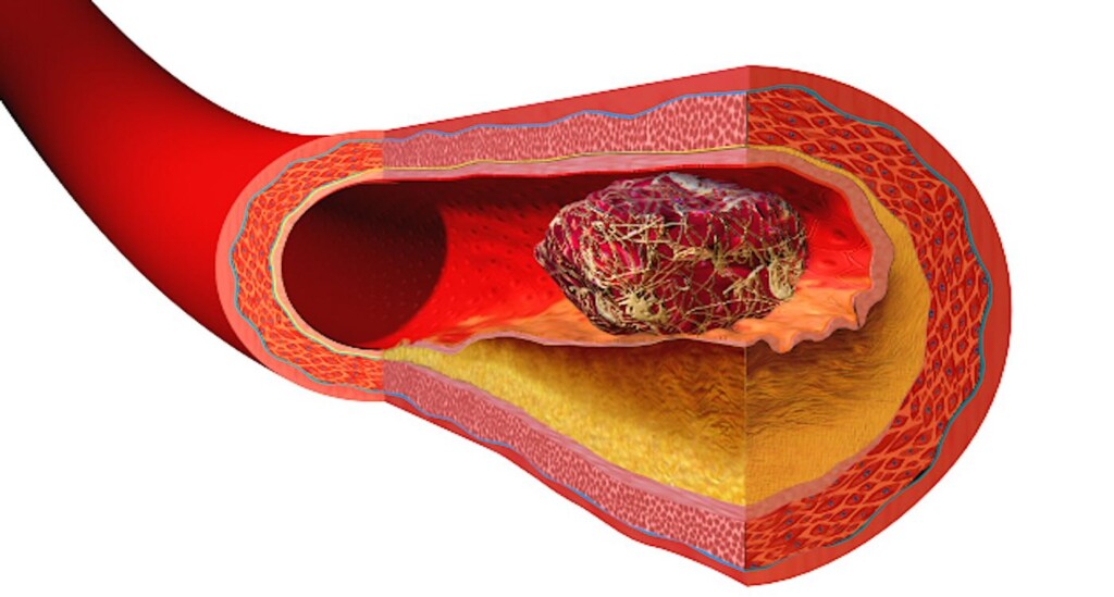
But they sometimes “react” and, in people with heart disease, they can form dangerous clots inside the arteries, causing heart attacks or cerebral vascular accidents.
“Plates play a crucial role in heart disease, in particular in coronary diseases, because they are directly involved in the formation of blood clots,” said the main study of the study, Dr. Kazutoshi Hirose, assistant professor at the University of Tokyo hospital.
To prevent dangerous clots, patients with coronary artery disease are often treated with anti-platform drugs. However, it is always difficult to assess with precision how these drugs work in each individual.
Professor Hirose and his team have developed a new system to monitor the plates while they are in motion, using a high -speed optical device and an AI.
“Like the traffic cameras capture each car on the road, our microscope captures thousands of images of blood cells in motion every second,” said the study co-author, Dr. Yuqi Zhou, professor of chemistry at the University of Tokyo, in a press release.
“We used an advanced device called Multiplexed Frequency Division Multiplex (FDM), which works as a super -speed camera that takes clear images of blood cells. We then use artificial intelligence to analyze these images. ”
Better than science fiction:: New electric bandages heal 30% faster than conventional dressings
“The AI can say if it looks at a single brochure – like a car – or a tuft of platelets, such as a traffic jam, or even a white blood cell that would hear – like a police car captured in the jam.”
The research team applied the technique to blood samples of more than 200 patients.
The results, published in the Journal Nature Communications, revealed that patients with acute coronary syndrome had more platelet aggregates than those with chronic symptoms – supporting the idea that technology can follow the risk of coagulation in real time.
The researchers said that one of the most important conclusions was that this technique can simply use blood taken from the arm, rather than heart arteries, and that it can provide almost the same information.
“It’s exciting because it makes the process much easier, safer and more practical,” said Dr. Hirose.
“As a general rule, if doctors want to understand what is happening in the coronary arteries, they must do invasive procedures, such as the insertion of a catheter through the wrist or groin to collect blood.”
BREAKTHROUGH:: Blood test which detects sepsis in 10 minutes by tightening blood cells – greeted as “the holy grail”
“Taking an ordinary blood sample from a vein in the arm can always provide significant information on platelet activity in the arteries.”
The team believes that technology will help doctors better personalize the treatment of heart disease.
“Just as some people need more or less an analgesic according to their body, we have found that people react differently to anti-platform drugs. In fact, some patients are affected by recurrent thrombosis and others suffer from recurrence of bleeding events even on the same anti-platia drugs.
Blood test for blows:: Researchers are developing a “revolutionary” blood test for the detection of strokes in the field: “Really transformer”
“Our technology can help doctors to see how the platelets of each individual behave in real time (and) IA can” see “the models beyond what the human eye can detect.”
The study also shows that even something as small as a blood cell can tell a great story about your health.
Share fantastic medical news with scientific geeks on social networks…


